Mammography
Mammography is a diagnostic imaging technique that uses the lowest possible dose of X-rays to visualize the tissue structure of the breast. This method allows for the detection of abnormalities in the fatty, glandular, and connective tissues of the breast, including early-stage breast cancer.
At Borstzorgklinieken, we have one of the latest mammography machines, known as Tomosynthesis or 3D mammography. The advantages of this state-of-the-art equipment includes the creation of a high-quality image with the lowest possible radiation dose, making abnormalities easier to detect and more visible, even in women with dense breast tissue. This is a valuable addition to the traditional 2D mammography.
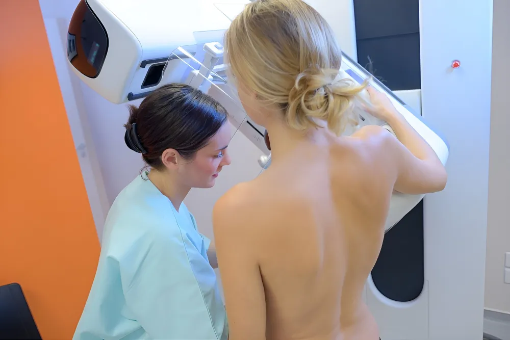
To make breast tissue visible and assess it accurately, some compression is applied to the breasts, and a picture is taken of both breasts. We have the "comfort compression plate" to minimize breast compression and optimize patient comfort during the examination. The examination takes less than 5 seconds per breast and is performed by our experienced mammography technologists, who will discuss with you how the examination can be conducted most comfortably. You can indicate how much pressure you can tolerate, which will reduce discomfort during the examination.
Pain and negative past experiences are often seen as barriers to getting a mammogram. Our mammography technologist specializes in addressing anxiety and will ask about your experiences. Based on your story, she will discuss how to successfully complete the examination, with your feelings and experience at the center of the process.
Mammography can also be performed if you have had breast surgery and/or breast implants, in the latter case without any risk to the implants themselves.
Following the mammography, our specialized breast radiologist will discuss the results with you. Sometimes, an additional biopsy or puncture may be necessary. The tissue collected in these procedures is sent for analysis to a pathologist, and the results are typically available within a few days.
If treatment is required, it will take place in a hospital. A multidisciplinary team will present the best treatment option(s) to you, and you will make a choice together.
Borstzorgklinieken collaborates with Antoni van Leeuwenhoek hospital, a specialized cancer hospital and research institute.
Timing of Mammography in the Menstrual Cycle
Since breast compression can sometimes be sensitive, we recommend scheduling the examination approximately 7 days after the start of your menstrual cycle. Your breasts are less sensitive during this time, making the examination more comfortable.
Pregnancy
During pregnancy, we typically do not perform mammography because unborn children are sensitive to X-rays, which is undesirable. It is advisable, however, to monitor the breasts during pregnancy if you have breast-related complaints. In such cases, we prefer using ultrasound.